
Intraoperative application of the TIVITA® Tissue © Stefan Straube
HSI. Hyperspectral Imaging.
Hyperspectral imaging: Imaging and visualization of comprehensive or complex chemical tissue properties.
HSI combines imaging, spectroscopy and tissue oximetry. It is used to image chemical properties with a high spectral resolution.
![]() Our hyperspectral camera systems bear the brand name TIVITA® and function according to the so-called pushbroom spectrometer principle. The chemical information of an object transported with the light is detected line by line. Thus, the measurement of a hyperspectral data cube requires a few seconds.
Our hyperspectral camera systems bear the brand name TIVITA® and function according to the so-called pushbroom spectrometer principle. The chemical information of an object transported with the light is detected line by line. Thus, the measurement of a hyperspectral data cube requires a few seconds.
The TIVITA® technology uses 100 wavelengths in the wavelength range from 500 to 1000 nm. This visual and near-infrared wavelength range contains various chemical information of the tissue. Among other things, the following physiological parameters are recorded:
- Oxygen saturation in superficial tissue layers
- perfusion in deeper tissue layers
- Hemoglobin content
- Water content
- Fat content
It would also be possible, based on the tissue-specific light spectra, to differentiate or classify different tissues and tissue changes by means of hyperspectral imaging algorithms. Such a classification usually takes place by means of artificial intelligence.
For the application and evaluation a broadband light source is required, which covers the entire wavelength range to be evaluated.
- Our hyperspectral camera systems work with imaging transmission spectrometers.
- The spectrometer unit consists of an entrance slit, two imaging optics, a holographic transmission grating and the area sensor. Via the input objective, the light remitted from the object passes through the input slit onto the first optics of the spectrometer.
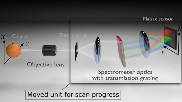
- The optics bundle the light and throw it onto the transmission grating, where it is split into individual wavelengths. Then it reaches the area sensor of the CMOS camera via a second optical system. Due to the design of the spectrometer unit, a spatial direction (width of the object as Y-axis) is detected.
- The second spatial direction (length of the object, X axis) results from the scan unit. It shifts the input slit and thus scans the object lengthwise within a few seconds. A third, spectral dimension is represented by the recorded wavelengths. This results in a 3D data cube (X [spatial dimension], Y [spatial dimension], λ [spectral dimension]).
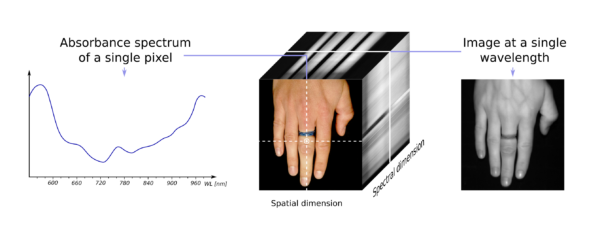
The 3D data cubes are the basis for the extraction of the chemical information and the various images. It is therefore possible to display the specific wavelength for each individual pixel in a generated image and use it for analysis or diagnostics.
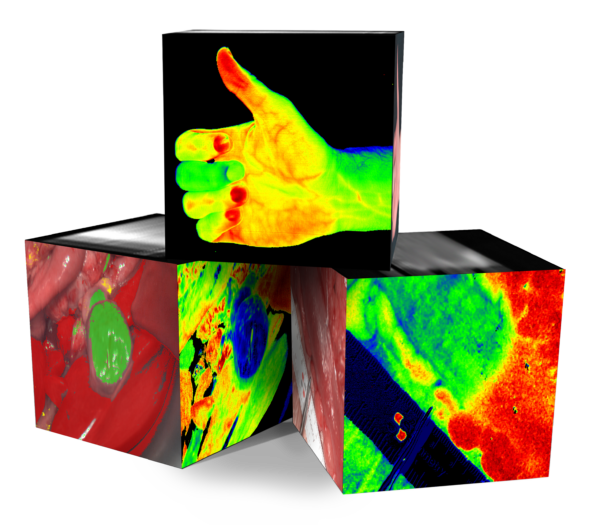
Various parameters can be calculated from the different wavelength ranges of the three-dimensional HSI cube and displayed in false-color images.
MSI. Multispectral Imaging.
Multispectral imaging: acquisition and live visualization of chemical tissue properties.
The information transported by the LED light is detected in just a few frames, which means that playback can be very fast and the technology is video-capable.
![]() Multispectral imaging offers the advantage of being video-capable due to good data availability, short measurement times and high resolution, thus enabling continuous live monitoring. Our multispectral camera systems carry the brand name MALYNA®.
Multispectral imaging offers the advantage of being video-capable due to good data availability, short measurement times and high resolution, thus enabling continuous live monitoring. Our multispectral camera systems carry the brand name MALYNA®.
All products of the MALYNA® series use some specific wavelengths in the range of 400 to 1000 nm. Thus, narrowband signals in characteristic ranges of the visible and near-infrared wavelength range are used to evaluate and display the parameters.
All products of the MALYNA® series use some specific wavelengths in the range of 400 to 1000 nm. These are provided individually one after the other by a multispectral high power LED illumination and recorded with a synchronized high resolution camera. To evaluate and display the parameters, the narrowband signals are analyzed in characteristic ranges using algorithms.
ICG imaging: acquisition and live visualization of tissue perfusion
![]() Indocyanine green (ICG) is one of the most widely used contrast agents in medicine. The associated imaging, which is based on visualization of the specific reflectance spectrum of this fluorescent dye, can be integrated into HSI and MSI imaging.
Indocyanine green (ICG) is one of the most widely used contrast agents in medicine. The associated imaging, which is based on visualization of the specific reflectance spectrum of this fluorescent dye, can be integrated into HSI and MSI imaging.
Image series: Highest quality for single images and image transmissions
![]()
![]()
![]() 4K sensor technology with continuous 4K transmission to the output medium, high-performance LED illumination and fast image transmission with minimum latency are quality features for an image section that enables precise and fatigue-free work on the patient.
4K sensor technology with continuous 4K transmission to the output medium, high-performance LED illumination and fast image transmission with minimum latency are quality features for an image section that enables precise and fatigue-free work on the patient.
Maximum miniaturization with chip-in-tip technology
![]() Due to the miniaturization of the individual components, the imaging systems can be integrated more flexibly and gently into everyday clinical practice. Flexible endoscopes in particular benefit from this development.
Due to the miniaturization of the individual components, the imaging systems can be integrated more flexibly and gently into everyday clinical practice. Flexible endoscopes in particular benefit from this development.
Computer-aided image data processing offers diagnostic support in everyday surgical practice
In the expectation of making physiological spectral data usable for tissue classification and later for specific diagnostics, a field of Computer Aided Detection and Computer Aided Diagnostics is growing. The detection of necrosis features is a first feature of the TIVITA® 2.0 Wound.