In weiteren Studien wird die HSI-Technologie als Methode zur objektiven Gewebebeurteilung in der Schilddrüsenchirurgie untersucht. Die Autofluoreszenzeigenschaft der Nebenschilddrüse bietet einen vielversprechenden Ansatz bei der Resektion der Nebenschilddrüse, diese intraoperativ zu lokalisieren und von Schilddrüsenknoten, Fettgewebe, Nervensträngen und Lymphknotengewebe zu unterscheiden.
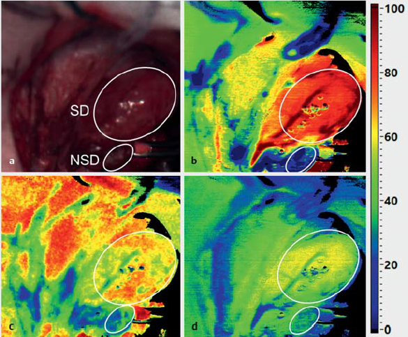
Erste Arbeiten mit der HSI Absorptionstechnik zur Identifikation der Nebenschilddrüse zeigen, dass insbesondere der Wassergehalt, die Oxygenierung und NIR-Perfusion in der Nebenschilddrüse geringer sind als in der Schilddrüse (Barberio et al. 2018, Gockel et al. 2020). Computer-assistierte Verfahren bieten sich daher für die automatische Gewebe-Diskriminierung an. Insbesondere die spektrale Analyse des Fluoreszenzlichtes eröffnet einzigartige Möglichkeiten, das Verfahren zu optimieren.