Klinische Anwendungen
Plastische Chirurgie / Verbrennungsmedizin
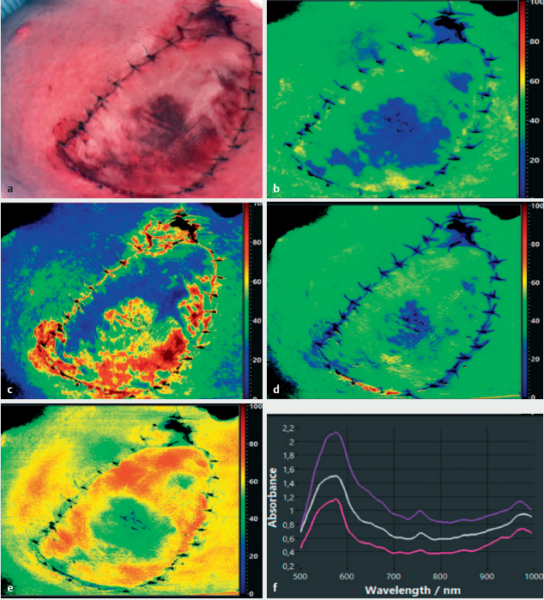
In der plastischen Chirurgie wird die HSI-Technologie u. a. für die dynamische und pathophysiologische Beurteilung von Lappenplastiken, Transplantaten sowie Brandwunden eingesetzt. Schlecht durchblutete Lappenplastiken sind bis zu 72 Stunden nach der Operation visuell klinisch nicht unbedingt auffällig. Zu dem Zeitpunkt, bei dem die schlechte Durchblutung erkannt werden kann, sind die Hautlappen jedoch in einem bereits schlechteren Zustand, damit schwieriger zu retten und verursachen einen höheren Revidierungsaufwand.
Mit der TIVITA® Tissue ist es dem Arzt/der Ärztin möglich, frühzeitig Oxygenierungs- und Perfusionsprobleme in Hautlappen, wie z. B. arterielle Insuffizienzen und venöse Stauungen, zu identifizieren und sofortige Korrekturmaßnahmen einzuleiten. Dadurch können Komplikationen reduziert und die Behandlungsergebnisse nach plastischen rekonstruktiven Operationen bedeutend verbessert werden.

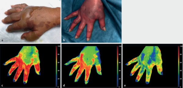
Des Weiteren bietet die HSI-Technologie ein nützliches Instrument zur objektiven Beurteilung und Bewertung von Verbrennungen und deren Heilungsprozessen sowie für die frühe Prognose der Verbrennungstiefe.
