MDSI. Medical Spectral Imaging.
Medical Spectral Imaging vereint Bildgebung, Spektroskopie und Gewebeoximetrie. Dynamisch. Nicht-invasiv. Kontaktlos.
MDSI. Medical Spectral Imaging.
MDSI. Die optische Messung physiologischer Gewebeparameter. Nicht-invasiv. Kontaktlos.
Die Perfusionsbildgebung erweitert als fünftes Bildgebungsverfahren das visuelle Sensorium in der Medizin.
Licht interagiert auf vielfältige Weise mit biologischem Gewebe. Beim Medical Spectral Imaging wird Gewebe mit Licht bestimmter Wellenlänge beleuchtet und das vom Gewebe remittierte Licht erfasst. Dieses remittierte Licht resultiert aus den spezifischen Absorptionseigenschaften der Gewebe bzw. Gewebebestandteile. In einigen Anwendungen kann auch die Fluoreszenz eine wichtige Rolle zur Auswertung spielen.
Mit den speziell entwickelten Kamerasystemen werden unterschiedliche Gewebeinformationen in verschiedenen Bereichen des elektromagnetischen Spektrums orts- und wellenlängenaufgelöst aufgenommen. Diese chemischen Informationen werden dann mittels Algorithmik in bildhafte Daten umgewandelt, um sie den Anwendern als Falschfarbenbilder verständlich darzustellen.

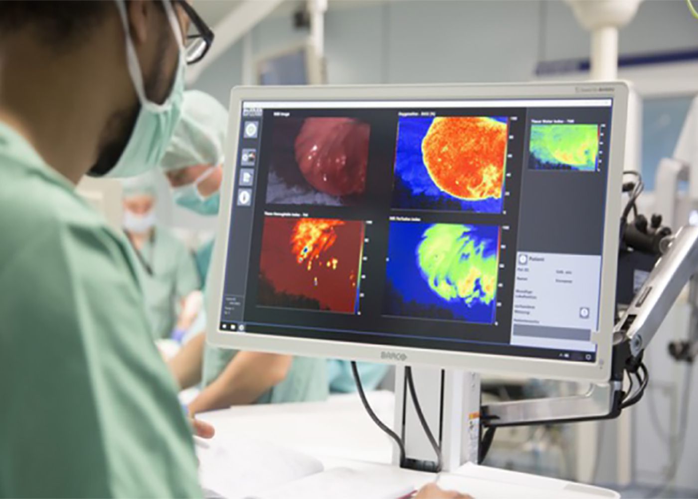
Intraoperative Anwendung der TIVITA® Tissue © Stefan Straube
HSI. Hyperspectral Imaging.
Hyperspektrale Bildgebung: Aufnahme und Darstellung umfassender oder komplexer chemischer Gewebeeigenschaften.
HSI vereint Bildgebung, Spektroskopie und Gewebeoximetrie. Es dient der bildhaften Aufnahme von chemischen Eigenschaften mit einer hohen spektralen Auflösung.
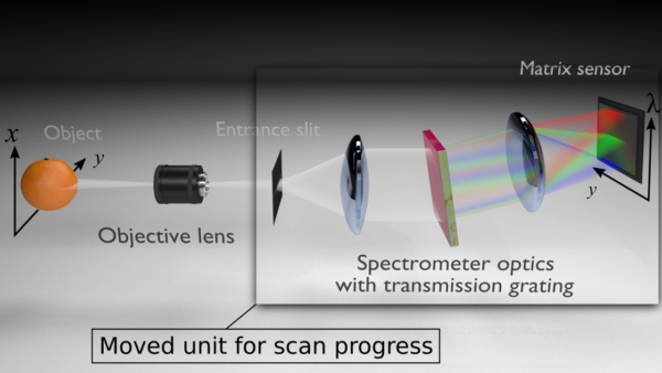
![]() Unsere hyperspektralen Kamerasysteme tragen den Markennamen TIVITA® und funktionieren nach dem sogenannten Pushbroom-Spektrometer-Prinzip. Die mit dem Licht transportierten chemischen Informationen eines Objektes werden zeilenweise detektiert. Somit benötigt die Messung eines hyperspektralen Datenwürfels einige Sekunden.
Unsere hyperspektralen Kamerasysteme tragen den Markennamen TIVITA® und funktionieren nach dem sogenannten Pushbroom-Spektrometer-Prinzip. Die mit dem Licht transportierten chemischen Informationen eines Objektes werden zeilenweise detektiert. Somit benötigt die Messung eines hyperspektralen Datenwürfels einige Sekunden.
Die TIVITA®-Technologie nutzt 100 Wellenlängen im Wellenlängenbereich von 500 bis 1000 nm. In diesem visuellen und nahinfraroten Wellenlängenbereich sind diverse chemische Informationen des Gewebes enthalten. So werden unter anderem folgende physiologische Parameter erfasst:
- Sauerstoffsättigung in oberflächlichen Gewebeschichten
- Perfusion in tieferen Gewebeschichten
- Hämoglobingehalt
- Wassergehalt
- Fettgehalt
Ebenso wäre es möglich, basierend auf den gewebespezifischen Lichtspektren, mittels hyperspektraler Bildgebung über Algorithmen unterschiedliche Gewebe und Gewebeveränderungen zu differenzieren bzw. zu klassifizieren. Eine solche Klassifizierung findet üblicherweise mittels künstlicher Intelligenz statt.
Für die Anwendung und Auswertung wird eine breitbandige Lichtquelle benötigt, welche den gesamten auszuwertenden Wellenlängenbereich abdeckt.
- Unsere hyperspektralen Kamerasysteme arbeiten mit bildgebenden Transmissionsspektrometern.
- Die Spektrometereinheit besteht aus einem Eingangsspalt, zwei Abbildungsoptiken, einem holographischen Transmissionsgitter und dem Flächensensor. Über das Eingangsobjektiv gelangt das vom Objekt remittierte Licht durch den Eingangsspalt auf die erste Optik des Spektrometers.

- Die Optik bündelt das Licht und wirft es auf das Transmissionsgitter, wo es in einzelne Wellenlängen zerlegt wird. Danach gelangt es über eine zweite Optik auf den Flächensensor der CMOS Kamera. Durch den Aufbau der Spektrometereinheit wird eine Raumrichtung (Breite des Objekts als Y-Achse) erfasst.
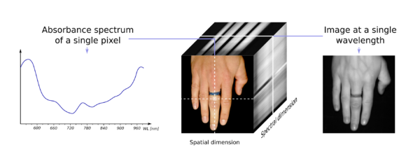
- Die zweite Raumrichtung (Länge des Objekts, X-Achse) ergibt sich über die Scaneinheit. Sie verschiebt den Eingangsspalt und scannt so das Objekt innerhalb weniger Sekunden der Länge nach ab. Eine dritte, spektrale Dimension stellen die aufgezeichneten Wellenlängen dar. Damit ergibt sich ein 3D-Datenwürfel (X [räumliche Dimension], Y [räumliche Dimension], λ [spektrale Dimension]).

Die 3D-Datenwürfel sind die Grundlage für die Extraktion der chemischen Informationen und der verschiedenen Bilder. Es ist also möglich, sich für jedes einzelne Pixel in einem generierten Bild die spezifische Wellenlänge darstellen zu lassen und zur Analyse bzw. Diagnostik heranzuziehen.

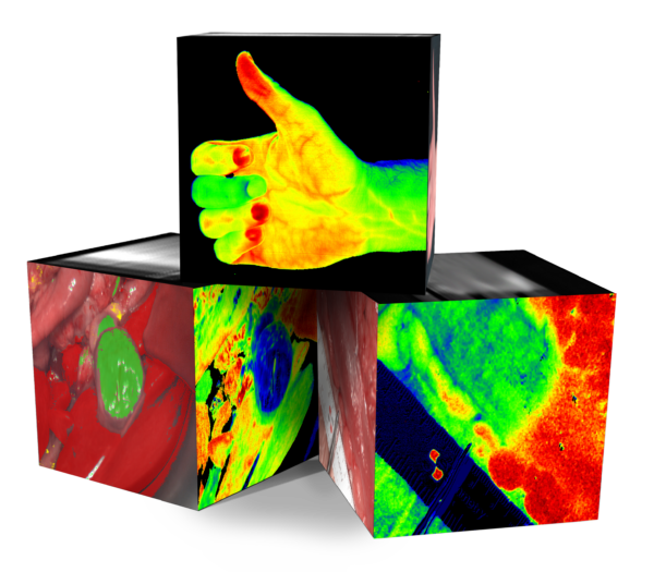
Aus den unterschiedlichen Wellenlängenbereichen des dreidimensionalen HSI-Cubes lassen sich verschiedene Parameter berechnen und in Falschfarbenbildern darstellen.
MSI. Multispectral Imaging.
Multispektrale Bildgebung: Aufnahme und Live-Darstellung chemischer Gewebeeigenschaften.
Die mit dem LED-Licht transportierten Informationen werden mit wenigen Bildern detektiert, wodurch die Wiedergabe sehr schnell erfolgen kann und die Technologie videofähig ist.
![]() Die multispektrale Bildgebung bietet den Vorteil, dass sie durch gute Datenverfügbarkeit, kurze Messzeiten und hohe Auflösung videofähig ist und dadurch ein kontinuierliches Live-Monitoring ermöglicht. Unsere multispektralen Kamerasysteme tragen den Markennamen MALYNA®.
Die multispektrale Bildgebung bietet den Vorteil, dass sie durch gute Datenverfügbarkeit, kurze Messzeiten und hohe Auflösung videofähig ist und dadurch ein kontinuierliches Live-Monitoring ermöglicht. Unsere multispektralen Kamerasysteme tragen den Markennamen MALYNA®.
Alle Produkte der MALYNA®-Serie nutzen einige spezifische Wellenlängen im Bereich von 400 bis 1000 nm. Zur Auswertung und Darstellung der Parameter werden somit schmalbandige Signale in charakteristischen Bereichen des sichtbaren und nahinfraroten Wellenlängenbereichs genutzt.
Alle Produkte der MALYNA®-Serie nutzen einige spezifische Wellenlängen im Bereich von 400 bis 1000 nm. Diese werden durch eine multispektrale High Power LED Beleuchtung einzeln nacheinander zur Verfügung gestellt und mit einer synchronisierten hochauflösenden Kamera aufgenommen. Zur Auswertung und Darstellung der Parameter werden die schmalbandigen Signale in charakteristischen Bereichen mit Hilfe von Algorithmen analysiert.
ICG-Bildgebung: Aufnahme und Live-Darstellung von Gewebeperfusion
![]() Indocyaningrün (ICG) ist eines der meistbenutzten Kontrastmittel in der Medizin. Die zugehörige Bildgebung, welche auf der Sichtbarmachung des spezifischen Reflektionsspektrums dieses Fluoreszenzfarbstoffs beruht, kann in die HSI- und MSI-Bildgebung integriert werden.
Indocyaningrün (ICG) ist eines der meistbenutzten Kontrastmittel in der Medizin. Die zugehörige Bildgebung, welche auf der Sichtbarmachung des spezifischen Reflektionsspektrums dieses Fluoreszenzfarbstoffs beruht, kann in die HSI- und MSI-Bildgebung integriert werden.
Bildstrecke: Höchste Qualität für Einzelbilder und Bildübertragungen
![]()
![]()
![]() 4K Sensorik mit durchgehender 4K Übertragung bis zum Ausgabemedium, Hochleistungs-LED-Beleuchtung und schnelle Bildübertragung mit geringster Latenz sind Qualitätsmerkmale für eine Bildstrecke, die präzises und ermüdungsfreies Arbeiten am Patienten ermöglicht.
4K Sensorik mit durchgehender 4K Übertragung bis zum Ausgabemedium, Hochleistungs-LED-Beleuchtung und schnelle Bildübertragung mit geringster Latenz sind Qualitätsmerkmale für eine Bildstrecke, die präzises und ermüdungsfreies Arbeiten am Patienten ermöglicht.
Maximale Miniaturisierung mit Chip-in-Tip Technologie
![]() Durch die Miniaturisierung der einzelnen Komponenten lassen sich die bildgebenden Systeme flexibler und schonender in den klinischen Alltag integrieren. Besonders die flexiblen Endoskope profitieren von dieser Entwicklung.
Durch die Miniaturisierung der einzelnen Komponenten lassen sich die bildgebenden Systeme flexibler und schonender in den klinischen Alltag integrieren. Besonders die flexiblen Endoskope profitieren von dieser Entwicklung.
Computergestützte Bilddatenverarbeitung bietet diagnostische Unterstützung im chirurgischen Alltag
In der Erwartung physiologische Spektraldaten für die Gewebeklassifizierung und später zur konkreten Diagnostik nutzbar zu machen, wächst ein Feld der Computer Aided Detection und Computer Aided Diagnostics. Die Erkennung von Nekrosemerkmalen ist ein erstes Feature bei der TIVITA® 2.0 Wound.
MDSI. Ausblick.
Was ist mit den Technologien noch möglich?
Mit zunehmendem technischem Fortschritt und medizinischem Verständnis der Daten werden sich komplett neue Möglichkeiten für die Diagnostik und damit für den klinischen Alltag ergeben.
Durch weitere Miniaturisierung von MDSI-Kameras können diese zukünftig z. B. als stereoskopische Kameras genutzt, als Chip-in-the-tip (CIT)-System für die starre oder flexible Endoskopie eingesetzt oder in robotische Assistenzsysteme integriert werden.
Die objektiven Daten könnten zudem als Grundlage für künstliche Intelligenz dienen, womit eine automatisierte Erkennung von Organen und Risikostrukturen keine Utopie mehr ist.
Ob HSI, MSI, ICG oder hochauflösende und miniaturisierte Bildstrecken – Wenn Sie Ideen, Fragen oder Anregungen haben, lassen Sie uns das gerne gemeinsam besprechen.