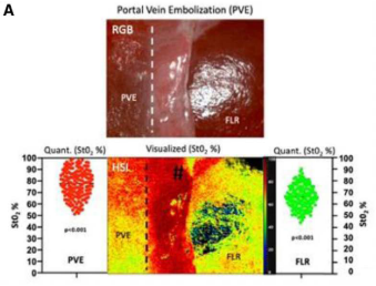
A huge milestone: The successful MDR certification

On 27.07.2023, it was a Thursday afternoon, the long-awaited news came from our Notified Body about the successful completion of the MDR certification process. After many invested hours of work, after numerous meetings and after seemingly endless waiting time, we can finally hold our MDR certificate in our hands.

We are overjoyed and very proud to have reached this existentially important milestone under difficult conditions. We now demonstrably meet the requirements of Regulation (EU) 2017/745 on medical devices and can thus from now on not only make our TIVITAproducts even safer and more efficient, but also develop new innovative products and make them available to patients and users on the medical device market.

A big thank you goes to the entire team at Diaspective for all the time and effort that went into this. We would like to explicitly mention the QM/RA team, without whom this successful implementation would not have been possible.

Many, many thanks!

HSI enables precise image-guided resection of the liver

Recently, Sucher et al. published in Annals of Surgery another valuable study on the application of HSI in liver surgery. This study investigated the effects of preoperative portal vein embolisation (PVE) and intraoperative vascular inflow control (VIC) on hepatic microcirculation and tissue oxygenation using HSI technology.

In a total of 58 patients with various liver diseases who underwent major liver resection, HSI technology enabled the visualisation of perfusion changes. In venous embolised liver segments, the effect of arterial hyperperfusion was impressively observed by significantly increased StO2 levels and NIR perfusion indices in the colour-coded HSI images. In contrast, total vessel occlusion by ligation of the portal vein and hepatic artery (VIC) resulted in an immediate and dramatic decrease in hepatic oxygenation and perfusion in the affected liver segments. The resulting optical borderline allowed a clear distinction between ischaemic and non-ischaemic tissue in all cases, irrespective of liver disease, thus facilitating precise image-guided parenchymal transection within the boundaries between perfused and non-perfused liver parenchyma.

Seeker et al. A


In addition, HSI made it possible to distinguish between different tumour entities in terms of their oxygen saturation, blood flow status and haemoglobin and water concentrations.

Seeker et al. B

We are very pleased about these impressive and trend-setting results and congratulate the authors on this high-quality publication!

Sucher E, Sucher R, Guice H, Schneeberger S, Brandacher G, Gockel I, Berg T, Seehofer D. Hyperspectral Evaluation of the Human Liver During Major Resection. Annals of Surgery. 2022;2:e169. doi: 10.1097/AS9.0000000000000169

https://journals.lww.com/aosopen/Fulltext/2022/06000/Hyperspectral_Evaluation_of_the_Human_Liver_During.18.aspx?context=LatestArticles

Professional exchange on developments in Medical Spectral Imaging (MDSI) at LASER World of PHOTONICS

This year, the LASER World of PHOTONICS trade fair is dedicating a separate platform within the "Biophotonics, Medical Applications, Optical Metrology and Imaging" forum to the topic "Biophotonics with DL meets Pathology, Point of Care and Laboratory Medicine / DGLM & LGT".
In addition to individual presentations, a panel discussion is also on the programme. There, Dr. Axel Kulcke, founder and CEO of Diaspective Vision GmbH, will share his many years of experience in the field of medical spectral imaging with the visitors.
Date: 26.04.2022, 9:00 -16:00 hrs - the panel discussion starts at 11:40 hrs
Location: LASER World of PHOTONICS, Munich Trade Fair Centre

Link: Application Panels: Topic Overview (world-of-photonics.com)

HSI is used to detect perfusion failure of flap grafts much earlier than by clinical assessment

The Mainz working group led by Dr Daniel Thiem achieved very impressive results in their clinical study on the monitoring of free flaps using HSI compared to clinical monitoring.

The studies on 63 patients with free flaps in the head and neck region showed that HSI is able to detect postoperative perfusion disturbances on average about 5 hours earlier than clinical monitoring with the same sensitivity as clinical assessment (100 %). These results are of great importance, as the early detection of perfusion disturbances is a decisive criterion for the success of treatment or the survival of the graft.

Thiem DGE, Römer P, Blatt S, Al-Nawas B, Kämmerer PW. New Approach to the Old Challenge of Free Flap Monitoring-Hyperspectral Imaging Outperforms Clinical Assessment by Earlier Detection of Perfusion Failure. J Pers Med. 2021;11:1101. doi: 10.3390/jpm111101

Thiem et al. 2021

Dot plot from Thiem et al. 2021 showing the duration (h) from flap insertion to clinical (red) and hyperspectral detection (green) of flap malfunction (Source: Thiem DGE et al. New Approach to the Old Challenge of Free Flap Monitoring-Hyperspectral Imaging Outperforms Clinical Assessment by Earlier Detection of Perfusion Failure. J Pers Med. 2021;11:1101).

MALYNA® live at the Medica/CompaMed in Düsseldorf

Compamed

At this year's Medica / CompaMed, which takes place from 15-18 November in Düsseldorf, we are represented by OmniVision, which is presenting its new OH08B high-performance CMOS imaging sensor in our prototype MALYNA® (Multispectral Imaging System) series. The new MALYNA® system combines live, ICG and multispectral perfusion imaging in a single camera system for the first time. OmniVision Technologies Inc. is a leading developer of advanced digital imaging solutions and is cooperating with us in the development of a new spectral camera system for endoscopes.

We are pleased to work with OmniVision to achieve greater confidence in diagnosis and decision-making through new approaches to medical imaging. Visit the OmniVision Technologies stand in Hall 14 / F40!

MALYNA MEDICA

Possibilities and perspectives of HSI in surgery

In the current review article by Barberio et al. the surgical audience gets a very nice overview of the different applications of hyperspectral imaging as an intraoperative imaging tool, the current limitations as well as the possible future direction of HSI for intraoperative guidance.

Based on a systematic literature review, the two current main areas of application for HSI, namely tissue detection (tumour detection and recognition of anatomical structures) and perfusion assessment in the context of various surgical procedures, are described.

The authors emphasise the great potential of hyperspectral imaging as an intraoperative decision support tool, as it has most of the characteristics of an ideal intraoperative imaging technology: HSI provides surgeons with reproducible and quantitative tissue information in a non-invasive, radiation-free, label-free and user-friendly manner.

Barberio M, Benedicenti S, Pizzicannella M, Felli E, Collins T, Jansen-Winkeln B, Marescaux J, Viola MG, Diana M. Intraoperative Guidance Using Hyperspectral Imaging: A Review for Surgeons. Diagnostics. 2021; 11(11):2066. doi.org/10.3390/diagnostics11112066.

Barberio et al.

First MALYNA® prototype on the test bench

Another milestone in the development of novel spectral camera systems for medical imaging has been reached! The first multispectral camera system of the MALYNA® series was delivered to one of our cooperation partners for extensive technical tests.

We are looking forward to the feedback in order to adapt the further development based on these results.

New publications on HSI

First HSI study in the field of pancreatic surgery

Several surgical disciplines have already used HSI, one of the leading new imaging systems, with demonstrable success to detect tissue perfusion. In a recent study by the Leipzig research group led by Ines Gockel, hyperspectral imaging has now been investigated for the first time as a helpful tool for assessing liver and gastric perfusion during pancreatoduodenectomy.

Source: Moulla Y, Buchloh DC, Köhler H, Rademacher S, Denecke T, Meyer HJ, Mehdorn M, Lange UG, Sucher R, Seehofer D, Jansen-Winkeln B, Gockel I. Hyperspectral Imaging (HSI)-A New Tool to Estimate the Perfusion of Upper Abdominal Organs during Pancreatoduodenectomy. Cancers. 2021;13:2846. doi: 10.3390/cancers13112846

https://www.mdpi.com/2072-6694/13/11/2846
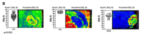
 

Video paper on precision surgery with HSI

A video paper by Jansen-Winkeln et al. impressively demonstrates how maximum precision can be achieved in rectal cancer by combining robotic surgery with intraoperative neuromonitoring and new imaging technologies such as HSI.

Source: Jansen-Winkeln B, Mehdorn M, Lange UG, Köhler H, Chalopin C, Gockel I. Precision Surgery In Rectal Resection With Hyperspectral And Fluorescence Imaging And Pelvic Intraoperative Neuromonitoring (With Video). Surg Technol Int. 2021;38:sti38/1383


HSI correlates with the conventional Modified Allen's Test (MAT)

In a study of 114 healthy patients, Heimes et al. show that HSI could serve as an additional method for assessing collateral blood flow in the hand prior to invasive procedures involving damage to or removal of the radial artery, since HSI can be used to reliably distinguish between perfusion and occlusion. In this respect, HSI shows a strong correlation to the previous gold standard, the modified Allen's Test (MAT). The authors emphasise the many advantages of HSI technology over visual assessment alone: HSI provides objective, reproducible results without interobserver error, can also be used by non-medical staff and provides visual and measurable feedback.

Heimes D, Becker P, Thiem DGE, Kuchen R, Kyyak S, Kämmerer PW. Is Hyperspectral Imaging Suitable for Assessing Collateral Circulation Prior to Radial Forearm Free Flap Harvesting? Comparison of Hyperspectral Imaging and Conventional Allen's Test. J Pers Med. 2021;11:531. doi: 10.3390/jpm11060531

https://www.mdpi.com/2075-4426/11/6/531

World novelty! TIVITA® Mini: First HSI camera for endoscopic and microscopic applications with CE marking!

After intensive work by our development and approval team, we are finally pleased to announce the registration of our TIVITA® Mini as a medical device for the European market.

With the TIVITA® Mini, the limitations of intraoperative HSI application have been overcome for the first time, as HSI is now also available for minimally invasive use. The TIVITA® Mini can be used as a flexible solution together with different optics.

We hope for a lot of interest and enthusiasm for this new intraoperative imaging method.

Optimisation of wound care in outpatient and inpatient facilities: TIVITA® Mobile approved as a medical device for the European market!

With the market launch of a hyperspectral hand-held camera, the TIVITA® Mobile, we complete the product portfolio of the TIVITA® series for the time being. This mobile version can be used independently of location for documentation purposes in wound therapy and also provide doctors and nurses with diagnosis-supporting information.