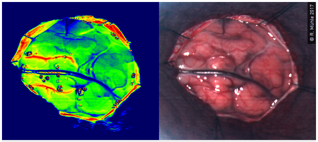
Zur Resektion von Hirntumoren werden präoperativ MRT-Aufnahmen gemacht, um den Tumor zu lokalisieren und anschließend die Planung des chirurgischen Eingriffs vornehmen zu können. Intraoperativ fehlen derzeit weitestgehend Hilfsmittel, den Tumor sicher zu lokalisieren.
Intraoperative Aufnahmen mit der TIVITA® Tissue während eines neurochirurgischen Eingriffs. HSI-Perfusionsdarstellung (links) und RGB-Kamerabild (rechts).
Ergebnisse mit der HSI-Technologie zeigen, dass es mit dieser Methode sehr wahrscheinlich möglich sein wird, den Tumor zu identifizieren. Die Arbeiten an diesem Thema werden in Kooperation mit dem Institut für Biomedizinische Technik der TU Dresden durchgeführt, welche bereits erste Veröffentlichungen dazu verfasst hat und einen kurzen Einblick auf Ihrer Homepage bietet.
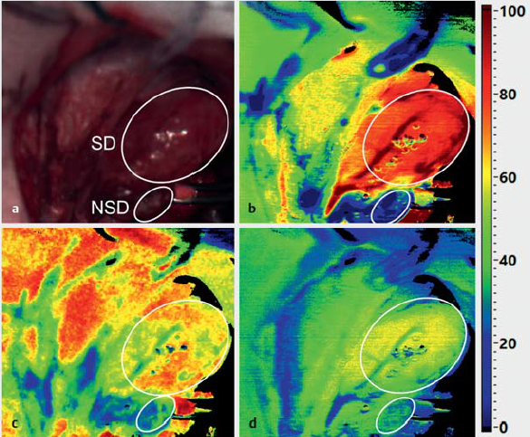
In weiteren Studien wird die HSI-Technologie als Methode zur objektiven Gewebebeurteilung in der Schilddrüsenchirurgie untersucht. Die Autofluoreszenzeigenschaft der Nebenschilddrüse bietet einen vielversprechenden Ansatz bei der Resektion der Nebenschilddrüse, diese intraoperativ zu lokalisieren und von Schilddrüsenknoten, Fettgewebe, Nervensträngen und Lymphknotengewebe zu unterscheiden.
Erste Arbeiten mit der HSI Absorptionstechnik zur Identifikation der Nebenschilddrüse zeigen, dass insbesondere der Wassergehalt, die Oxygenierung und NIR-Perfusion in der Nebenschilddrüse geringer sind als in der Schilddrüse (Barberio et al. 2018, Gockel et al. 2020). Computer-assistierte Verfahren bieten sich daher für die automatische Gewebe-Diskriminierung an. Insbesondere die spektrale Analyse des Fluoreszenzlichtes eröffnet einzigartige Möglichkeiten, das Verfahren zu optimieren.
RGB-Bild und HSI-Aufnahmen einer Schilddrüse (SD) und Nebenschilddrüse (NSD) mit deutlich sichtbar geringeren Werten für die Nebenschilddrüse. NIR-Perfusionsindex (b), StO2(c), Wasser-Index (d). (Bildquelle: Gockel I et al. Hyperspektral-Imaging (HSI) – Eine verlässliche Gewebedifferenzierung? Zentralbl Chir 2020; 145:125-129).
Neben Faktoren wie Alter und Gesundheitszustand des Spenders trägt die Organkonservierung entscheidend zur Funktionalität eines Organs und somit zum Erfolg der Transplantation bei.
In den letzten Jahren gab es diesbezüglich viele Bemühungen, die Organqualität zu verbessern. Im Vergleich zur klassischen Kryokonservierung, die meist aufgrund der kühlen Lagerung, Wiedererwärmung und Durchblutung im Empfänger mit Ischämie/Reperfusionschäden einhergeht, kann die (normotherme) Organperfusion und -konservierung durch dynamische Flussprinzipien (Maschinenperfusion) die Organqualität erheblich verbessern.
Mithilfe der TIVITA® könnte die Organqualität effizient, mit geringem Zeitaufwand und nicht-invasiv untersucht werden. Zusätzlich zur prä-operativen Analyse der Viabilität des Organs, kann die Organlagerung und -behandlung sowie der kurzfristige Erfolg der Transplantation am Ende der Operation (Kontrolle der Gewebeperfusion) anhand von Parametern zur Gewebeoxygenierung und Durchblutung beurteilt werden. Das heißt, die ChirurgInnen können bereits vor der OP objektiv beurteilen, ob das Organ zur Transplantation geeignet ist oder nicht. Zudem ist es ihm/ihr möglich, direkt nach der Transplantation zu überprüfen, ob die Blutgefäße richtig vernäht wurden und das Organ ausreichend mit Blut versorgt wird.
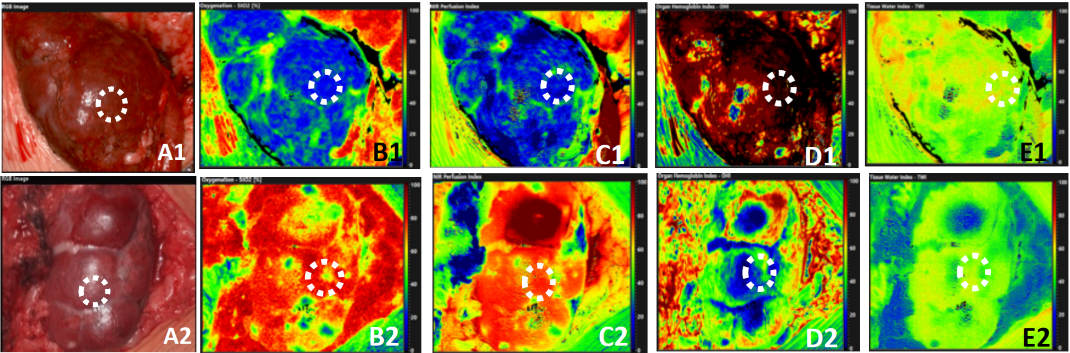
In einer aktuellen Studie von Sucher et al. (2020) konnte intraoperativ mittels HSI erfolgreich die Lebens- und Leistungsfähigkeit des Nierenparenchyms und des Harnleiters von Nierentransplantaten objektiv beurteilt und eine verzögerte Transplantatfunktion vorhergesagt werden.
Intraoperativ aufgenommene (A) RGB- und Falschfarben-Hyperspektralbilder für (B) Oxygenierung (StO2), (C) Perfusion (NIR Perfusion Index), (D) Hämoglobingehalt (OHI) und (E) Wassergehalt (TWI), mit Region of Interest (ROI)-Markern innerhalb des Parenchyms von Nieren-Allotransplantaten mit DGF (A1-E1) oder ohne DGF (A2-E2) zum Zeitpunkt der Transplantation (Quelle: Sucher R et al. Hyperspectral Imaging (HSI) of Human Kidney Allografts. Ann Surg. 2020 Nov 13.)
Die Methode der Organkonservierung mittels Maschinenperfusion wird aktuell in Leipzig und Innsbruck erforscht. In diesen Untersuchungen dient die TIVITA® als Monitoring-Instrument zur Beurteilung der Organqualität vor der Transplantation bzw. bei der Organbehandlung während der Maschinenperfusion.
Auch in der Dermatologie steht die Optimierung der Versorgung akuter und chronischer Wunden im Fokus vieler klinischer Studien. Neben der Wundreinigung stellen moderne bioaktive Wundauflagen sowie kaltes Atmosphärendruckplasma innovative Behandlungsoptionen dar. Großes Ziel in diesem Fachbereich ist es zudem, mittels der hyperspektralen Kameratechnik und künstlicher Intelligenz, Hautkrebs nicht-invasiv und sicher zu diagnostizieren.
HSI-Aufnahmen einer Wunde vor, direkt nach und 10 min nach Behandlung mit kaltem Atmosphärendruckplasma. Obere Reihe: StO2, untere Reihe: NIR-Perfusionsindex. Vor der Behandlung ist die Perfusion in oberen und tieferen Schichten im Wundbereich deutlich reduzierter als in der Umgebung. Durch die Behandlung mit kaltem Atmosphärendruckplasma steigt die Perfusion im Wundbereich signifikant an. (Bildquelle: Daeschlein G et al. Hyperspectral imaging: innovative diagnostics to visualize hemodynamic effects of cold plasma in wound therapy. Biomed Tech (Berl). 2018 Oct 25;63(5):603-608)
Im Rahmen einer Pilotstudie der Arbeitsgruppe um Prof. Emmert (Universitätsmedizin Rostock) wird aktuell überprüft, ob die Therapie mit kaltem Atmosphärendruckplasma den Heilungsverlauf von Spalthautentnahmestellen günstig beeinflusst und möglichen Komplikationen wie Infektionen vorbeugt. Hierbei werden der Verlauf der Wundheilung, u. a. durch die hyperspektrale Technologie, dokumentiert und so verschiedene Wundtherapien objektiv miteinander verglichen.
In einer weiteren Studie aus Greifswald wird die TIVITA eingesetzt, um den Wundheilungsverlauf von Spalthautentnahmestellen am Oberschenkel zu überwachen, welche mit einem Wundgel behandelt wurden.
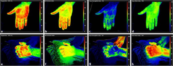
Besonders bei kritisch kranken Patienten sind mikrozirkulatorische Veränderungen mit einer erhöhten Mortalität und Morbidität verbunden. Daher ist es in der Intensiv- und Notfallmedizin besonders wichtig, neben der Makrozirkulation, auch die Gewebeperfusion und Ödembildung zu monitoren, um die negativen Auswirkungen einer hämodynamischen Inkohärenz zu reduzieren.
Hier stellt das Hyperspektral-Imaging eine vielversprechende Methode dar, um bettseitig Veränderungen der Mikrozirkulation, Oxygenierung und des Wassergehalts der Haut zu monitoren. Probleme wie Medikamentendosisfehler, Kreislaufstörungen, pulmonale Komplikationen oder ineffiziente Sauerstofftherapien könnten somit frühzeitig erkannt werden. Der Gewebe-Wasser-Index, als Parameter eines Gewebe- oder Darmödems, kann z. B. indirekte Rückschlüsse auf die anästhesiologische Steuerung der intraoperativen Volumentherapie zulassen.
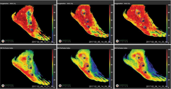
HSI-Aufnahmen der rechten Hand eines gesunden Probanden (a–d) und eines Patienten mit septischem Schock (e–h). Die Gewebeperfusion und-oxygenierung liegen im Bereich des gesunden Probanden, jedoch zeigen hohe TWI-Werte (h) ein ausgeprägtes Gewebeödem als mögliche Folge der Flüssigkeitstherapie und des Kapillarlecks. (Bildquelle: HySpI-ICU-Studie; Dietrich M et al. Bedside hyperspectral imaging for the evaluation of microcirculatory alterations in perioperative intensive care medicine: a study protocol for an observational clinical pilot study (HySpI-ICU). BMJ Open. 2020 Sep 17;10(9):e035742)
Dieses Beispiel zeigt sehr eindrucksvoll, die Fähigkeit der hyperspektralen Bildgebung mikrozirkulatorische/ödematöse Veränderungen in kritisch kranken Patienten bettseitig nachzuweisen. Somit kann frühzeitig die Therapie angepasst werden.
Die beiden ersten Prototypen unserer neuen TIVITA® 2.0 sind seit vier Wochen im praktischen Test in zwei Kliniken, mit denen wir bereits seit Jahren eng und fruchtbar zusammenarbeiten.
Ziel ist es, die Usability für das fertige Produkt zu überprüfen und schließlich zu optimieren, damit die zukünftigen Kunden ein Gerät erhalten, welches sie gerne zur Unterstützung ihrer täglichen Arbeit nutzen. Natürlich wird die Kamera im derzeitigen Stadium nicht am Patienten eingesetzt, sondern an Resektaten verwendet, um einen möglichst realen, klinischen Einsatz zu simulieren.
Wir freuen uns auf das kommende Feedback, um die TIVITA® 2.0 für Sie noch besser zu machen!
Um zukünftig das Medical Spectral Imaging für verschiedenste medizinische Anwendungen verfügbar zu machen, investieren wir stetig in Forschungs- und Entwicklungsaktivitäten.
Dabei konzentrieren wir uns auf die kontinuierliche Weiterentwicklung des bestehenden Produktportfolios und die Entwicklung neuer Produkte und Produktplattformen.
Wir bieten zuverlässige Unterstützung bei Ihrem Forschungsvorhaben.
Sie haben Ideen für neue medizinische Anwendungsgebiete oder Interesse an gemeinsamen Forschungsprojekten? Wir stehen jederzeit für einen Ideenaustausch zur Verfügung und diskutieren gerne mit Ihnen über Kooperationsprojekte oder individuelle Lösungsmöglichkeiten für Ihre Anwendung!
Wir sind zudem sehr daran interessiert, klinische Studien zu unterstützen, die sich auf die bildgebende nicht-invasive Gewebespektroskopie und Perfusionsanalyse konzentrieren.
Lassen Sie uns gemeinsam etwas bewegen.
Sie haben eine Produktidee im Bereich der medizinischen Spektralbildgebung und suchen nach der passenden Lösung? Gern beraten und unterstützen wir Sie in gemeinsamen Entwicklungsprojekten.
Gestalten Sie die Zukunft der spektralen Bildgebung mit.
Bei Diaspective Vision erwarten Sie herausfordernde Aufgaben und vielfältige Entwicklungschancen – unabhängig von Ihrem individuellen Einstiegslevel. Bewegen Sie gemeinsam mit uns Großes und werden Sie ein Teil unserer Vision!
Initiativbewerbungen können Sie gerne jederzeit an uns richten. Senden Sie Ihre Bewerbungsunterlagen an: Amadeus Holmer
Angebote für Studenten
Wir bieten Studierenden Einblicke in alle Bereiche eines Medizintechnikunternehmens. Bei uns erwarten Sie herausfordernde Aufgaben in einem dynamischen Arbeitsumfeld, bei denen Sie Ihre theoretischen Kenntnisse in die Praxis umsetzen können.
Absolvieren Sie bei uns ein Praktikum, übernehmen Sie eine Werkstudententätigkeit oder verfassen Sie bei Diaspective Vision Ihre Abschlussarbeit. Insbesondere im technischen Bereich stehen Ihnen diverse Themen im Bereich Forschung und Entwicklung, Konstruktion, IT und unserer Medizinproduktgruppen zur Verfügung.
Die Diaspective Vision GmbH beschäftigt sich schwerpunktmäßig mit der Entwicklung und Produktion von Kamerasystemen im Bereich Medical Spectral Imaging. Unsere Vertriebspartner sorgen für die Verbreitung unserer zugelassenen Medizinprodukte im klinischen Umfeld.
Kontaktieren Sie uns auch gerne direkt mit Ihrer Anfrage. Wir beantworten Ihre Fragen und helfen bei der Vermittlung der zuständigen Vertriebspartner.
Protex Healthcare GmbH
Die Protex Healthcare GmbH vertreibt unsere Produkte außerhalb der DACH-Region in den Fachgebieten:
Um dir ein optimales Erlebnis zu bieten, verwenden wir Technologien wie Cookies, um Geräteinformationen zu speichern und/oder darauf zuzugreifen. Wenn du diesen Technologien zustimmst, können wir Daten wie das Surfverhalten oder eindeutige IDs auf dieser Website verarbeiten. Wenn du deine Zustimmung nicht erteilst oder zurückziehst, können bestimmte Merkmale und Funktionen beeinträchtigt werden.
Funktional
Immer aktiv
Die technische Speicherung oder der Zugang ist unbedingt erforderlich für den rechtmäßigen Zweck, die Nutzung eines bestimmten Dienstes zu ermöglichen, der vom Teilnehmer oder Nutzer ausdrücklich gewünscht wird, oder für den alleinigen Zweck, die Übertragung einer Nachricht über ein elektronisches Kommunikationsnetz durchzuführen.
Vorlieben
Die technische Speicherung oder der Zugriff ist für den rechtmäßigen Zweck der Speicherung von Präferenzen erforderlich, die nicht vom Abonnenten oder Benutzer angefordert wurden.
Statistiken
Die technische Speicherung oder der Zugriff, der ausschließlich zu statistischen Zwecken erfolgt.Die technische Speicherung oder der Zugriff, der ausschließlich zu anonymen statistischen Zwecken verwendet wird. Ohne eine Vorladung, die freiwillige Zustimmung deines Internetdienstanbieters oder zusätzliche Aufzeichnungen von Dritten können die zu diesem Zweck gespeicherten oder abgerufenen Informationen allein in der Regel nicht dazu verwendet werden, dich zu identifizieren.
Marketing
Die technische Speicherung oder der Zugriff ist erforderlich, um Nutzerprofile zu erstellen, um Werbung zu versenden oder um den Nutzer auf einer Website oder über mehrere Websites hinweg zu ähnlichen Marketingzwecken zu verfolgen.