Den Anwendern werden so zusätzliche diagnoseunterstützende Informationen zur Verfügung gestellt, welche mit dem menschlichen Auge nicht sichtbar sind. Durch die Erweiterung des Sichtfeldes des Chirurgen und die Unterstützung bei der Erkennung von anatomischen Risikostrukturen und pathologischen Veränderungen kann bei komplexen Operationen das klinische Outcome für die Patienten potentiell verbessert werden.
Die Grundlage für die Entwicklung der TIVITA® Mini bildet die TIVITA® Tissue, welche die Technologie der hyperspektralen Bildgebung u. a. bereits für offen-chirurgische Anwendungen etabliert hat und erfolgreich intraoperativ, z. B. zur Beurteilung von Anastomosen und zur präzisen Festlegung von Resektionspositionen, eingesetzt wird.
Die TIVITA® Mini kann in allen Fachrichtungen angewendet werden, in denen die minimal-invasive Chirurgie zum Einsatz kommt, zum Beispiel in der:
Die Vorteile
- Darstellung von Durchblutungs- und Substanzparametern zur Erkennung von anatomischen Risikostrukturen
- Kontaktlose Bildgebung
- Nicht-invasiv, da keine Farbstoffe notwendig sind
- Ungefährlich, da keine ionisierende Strahlung
- Einfache Bedienung durch intuitive Software
- Keine Vorbereitungszeit notwendig – minimaler Einfluss auf den Workflow
- Schnelle und repetitive Messungen
- Objektiv durch stabile Algorithmen
- Kosteneffektiv
Die TIVITA® Mini wird als Gesamtsystem angeboten, bestehend aus den Kernkomponenten: endoskopische HD-Kamera für Live-Video und HSI-Aufnahmen, Imaging Center (breitbandige LED-Beleuchtungseinheit, leistungsstarker Rechner) inklusive Anwendungssoftware, zwei Monitore, endoskopische Optik und optional mit Gerätewagen und Fußschalter.
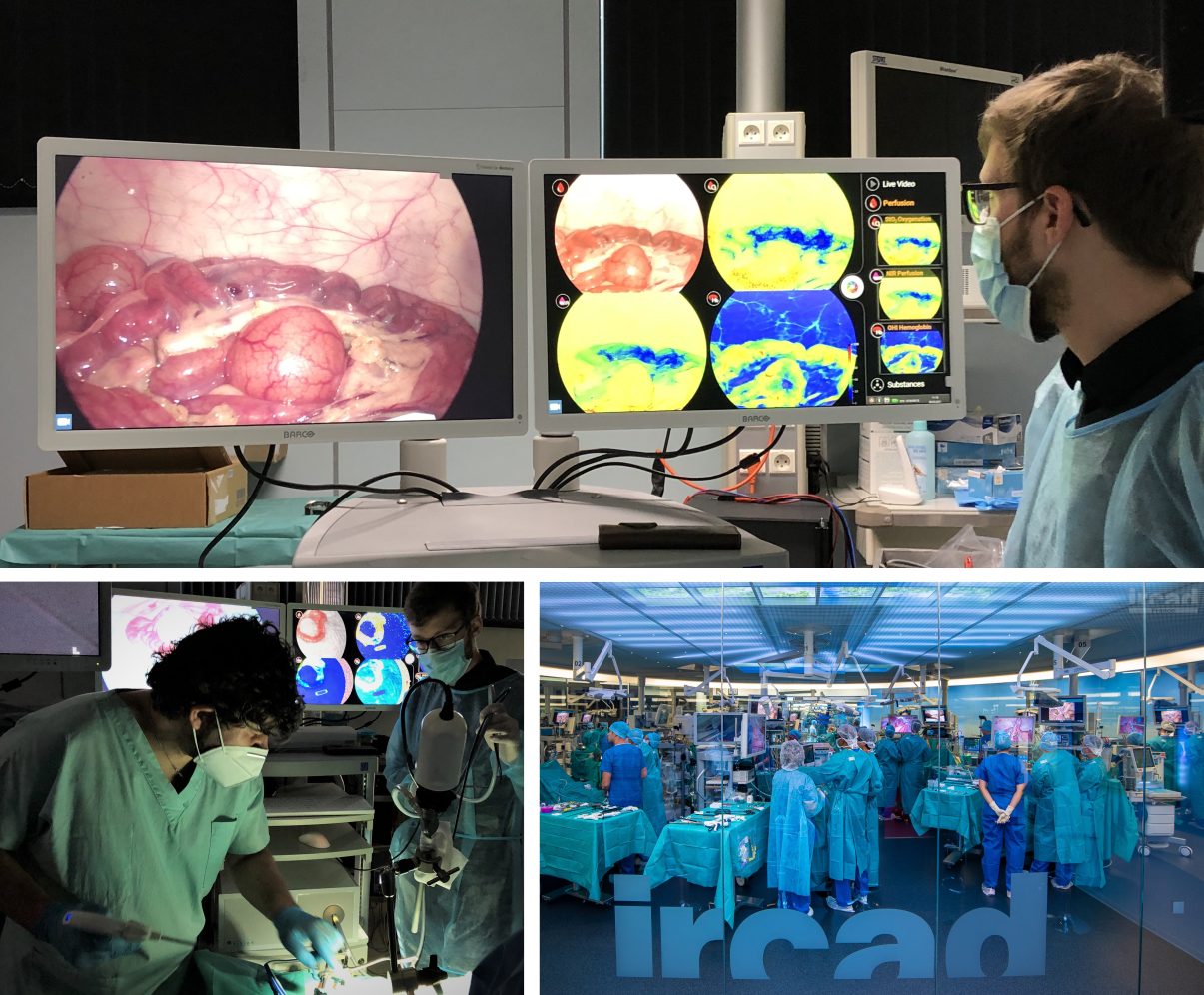
Dank verschiedener Kamerasensoren ist es möglich, dauerhaft ein Farbvideo zu sehen und gleichzeitig beliebig viele HSI-Aufnahmen zu machen, welche auf dem zweiten Monitor dargestellt werden. Mit nur einer hyperspektralen Aufnahme durch die TIVITA® Mini werden Ihnen verschiedene Durchblutungs- und Substanzparameter dargestellt. Dazu gehören:
- Gewebe-Sauerstoffsättigung (StO2 [%])
- Organ-Hämoglobin-Index (OHI [Indexwert])
- NIR-Perfusion-Index (NIR [Indexwert])
- Tissue-Water-Index (TWI [Indexwert])
- Tissue-Lipid-Index (TLI [Indexwert])
In verschiedenen Studien wird die hyperspektrale Bildgebung mit der ICG-Fluoreszenz-Bildgebungstechnik zur intraoperativen Perfusionskontrolle verglichen. Bisherige Untersuchungen zeigen eindeutig, dass HSI und FI-ICG vergleichbare Bildgebungsverfahren sind, zuverlässige Prädiktoren für eine angemessene Durchblutung darstellen und komplementär eingesetzt werden können.
Die HSI bietet gegenüber der ICG-Bildgebung entscheidende Vorteile.
Im Vergleich zur HSI gehen Fluoreszenz-basierte Technologien mit einigen Risiken für die Patienten einher, da im Gegensatz zur nicht-invasiven HSI die Injektion eines Fluoreszenzfarbstoffes notwendig ist. So sind phototoxische Effekte und Überempfindlichkeitsreaktionen beschrieben. Nachteilig ist ebenso, dass entzündliche Veränderungen, Abszesse und nekrotische Areale in falsch positiven Fluoreszenzen resultieren. Zudem ist die Qualität des Signals stark von der Gewebeart abhängig und kann zusätzlich negativ durch den Effekt des „Photo-bleaching“ beeinflusst werden.
Außerdem ist die Verwendung des TIVITA® Mini Systems kostengünstiger, da allein der Materialeinsatz von 25 mg ICG ca. 80 € kostet [Baiocchi GL, Diana M, Boni L. Indocyanine green-based fluorescence imaging in visceral and hepatobiliary and pancreatic surgery: State of the art and future directions. World journal of gastroenterology, 2018, 24. Jg., Nr. 27, S. 2921] – diese laufenden Kosten entfallen für HSI komplett. Diese Einsparung entspricht einem Wert von 80.000 – 240.000 € in fünf Jahren, wenn ein bis drei Einsätze pro Tag bei 200 Arbeitstagen im Jahr angenommen werden. Weitere Vorteile der HSI sind die kurze Vorbereitungs- und Messzeit, Reproduzierbarkeit und Wiederholbarkeit der Aufnahmen, unabhängig von der Verteilung eines Farbstoffes im Organismus.
Hier finden Sie Ihren Vertriebspartner und eine Liste mit wichtiger Literatur.